Next Step For Tissue Engineered Equine Cartilage Will be Evaluation in Live Horses
Equine Guelph
by: Jackie Bellamy-Zions
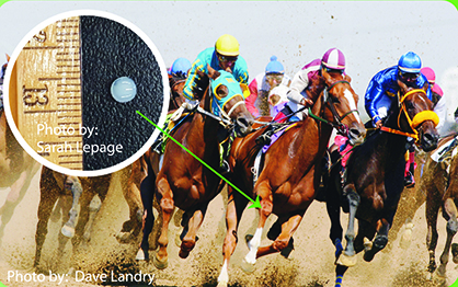
“It’s approximately four millimeters in diameter,” exclaimed Ontario Veterinary College researcher, Thomas Koch, unable to contain his excitement. The tiny disk of equine cartilage, manufactured in the OVC lab, is full of potential.
A cartilage injury can mean the end of an athlete’s career. Damaged joint cartilage does not repair on its own and often leads to early osteoarthritis.
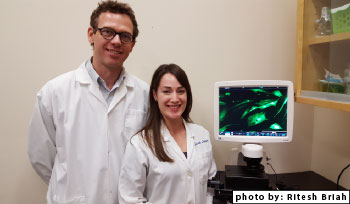
Great progress has been made, in Koch’s lab, by PhD student Sarah Lepage, in collaboration with Dr. Rita Kandel from the University of Toronto. They are putting together a protocol for making tissue engineered cartilage constructs. The next step will be evaluating them in live horses. “We are standing on the shoulders of research pioneered by Dr. Mark Hurtig and Kandel,” says Koch, crediting the developers of the mosaic grafting technique of bone and cartilage for sheep and horses. The graft keeps transplanted tissue in place for the healing process to begin.
Now that the equine cartilage disk is a reality, the question becomes “how will it hold up to the larger forces exerted in a horse?” There is further refinement and testing to be done before the first live trial on an equine, potentially by 2017. This topic is sure to be of great interest to human medicine as well, given the similar cartilage thickness and great athletic ability of the horse.
Koch says, “It is very exciting and satisfying to be this close to testing the tissue construct in a horse.” Ten years of exploratory work, finding the best possible cells, carrier substances and protocols are paving the way to revolutionary methods of treating cartilage damage with cell-based therapies.
The Best Cells
Mesenchymal Stromal Cells (MSC’s) are the building blocks of bone, cartilage, fat, muscle and tendon. When Koch began isolating MSC’s in 2005 he discovered they could be sourced from umbilical cord blood of newly born foals.
His team discovered isolating MSC’s from jugular blood was also possible but not as reliable. Cord blood cells taken at birth are younger and possess superior capabilities of dividing and creating different types of tissues. Koch explains, “They are easy to make into cartilage cells and better at it than other alternatives such as using cells from bone marrow, adipose tissue, and equine-induced pluripotent stem cell (iPSC).”
iPSC cells were explored by PhD Candidate, Sarah Lepage, in a collaborative study with Drs. Rita Kandel, Kristina Nagy, and Andraas Nagy from the University of Toronto. Desirable for their expandability and differentiation capacity, Lepage compared iPSC cells to MSC cells with the initial goal of making cartilage cells from the iPSC cells. With the cell lines they had available, they were not able to make it happen but Lepage was able to make the iPSC’s into a cell similar to MSC. If they gain access to more iPSC cells in the future, they now have a method to differentiate them into an MSC type cell for further evaluation. The technology behind making iPSC cells remains a point of interest.
After extensive investigation of the sources mentioned above, MSC’s from cord blood proved to be the cell of choice for Koch’s research.
Promising Results
Injection of cord blood MSC’s into joints of research horses has proven safe in multiple studies.
In collaboration with Dr. Judith Koenig, Clinical Studies at OVC, former PhD Candidate, Dr. Lynn Williams, made significant contributions to research, performing a live study injecting lipopolysaccharide (LPS), which induces a temporary inflammatory response. He performed injections with and without MSC’s. In the blinded study, results showed less inflammation in the equine joints for the injections which included MSC’s.
Williams also studied how injected stem cells suppress lymphocytes, a modulatory function of the immune system. In a comparison study, he discovered there is no difference between using frozen cells (freshly thawed out of liquid nitrogen), and those that have been thawed and allowed to adjust for a week in the lab. The clinical applications of having viable cells for treatment right after thawing from liquid nitrogen eliminates the need for a cell culture facility and expedites treatment.
Williams also played a role in determining the best carrier solution to transport the MSC for clinical use in collaboration with a U.S. company, BioLife Solutions. Stem cells, shipped chilled in a carrier solution, need to maintain viability. Carrier solutions containing biologics such as: autologous serum from the horse itself, bone marrow supernatant or platelet rich plasma (PRP) could prove detrimental to the cells and make analyzing results more difficult.
Keith Russell, a PhD Candidate in the Koch lab, last year showed that platelet lysate derived from PRP is in fact detrimental to MSC growth in vitro. Williams tested MSC cells injected into joints with a carrier solution, HypoThermosol, which is free from biologics (no proteins or serum) and compared results to MSC cells injected using saline. No difference in response was noted between the HypoThermosol and saline groups. The commercial carrier solution, free from biologics, is now used when testing MSC in live horses. “Often stem cells are combined with other biologics, but the absence of biologics in this formula allows our researchers to attribute the results of stem cell treatments directly to MSCs,” emphasizes Koch.
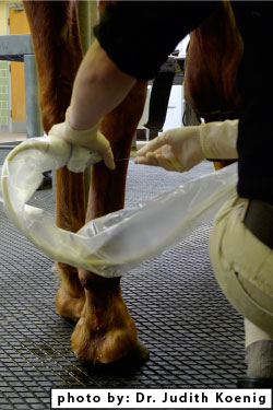
More success stories were recorded in the last few years when three client owned horses with tendon damage returned back to work, following cord blood MSC injection. No adverse reactions were noted and all three were treated with MSC cord blood acquired from another horse. In the one horse, saline was used as the carrier and in the other two HypoThermosol was used. This work was also done in collaboration with Dr. Judith Koenig.
One case was a breeding stallion, with such a severe tendon injury, he was unable to mount the phantom for collection. He became a candidate for stem cell therapy after previous surgery and extensive rehabilitation proved unsuccessful. He returned to work four months later, after two MSC cord blood injections, given one month apart. The imaging specialist was very surprised when the ultrasound revealed incredibly fast healing of the tissue including fiber alignment and filling of the defect. Drs. Koch and Koenig are now recruiting horses with tendon injuries for a controlled study to determine efficacy of the stem cells in tendon healing.
Koch explains, “The effect seen in the tendons after MSC stem cell therapy is most likely due to the cells immunomodulatory properties.” Meaning they are capable of modifying or regulating one or more immune functions.
The MSC cells are:
1) influencing and reducing inflammation at the site
2) providing a micro-environment that is more conducive to healing
3) secreting different factors that are influencing the immune system
Bright Future and More Live Studies on the Horizon
“We are interested in exploring how these cells are able to modulate the immune system,” says Koch, “as well as evaluating MSCs for their capacity to become different cell types, and in particular cartilage.” Investigating the secretory functions is a future topic of study for the OVC lab, with a PhD student ready to assist. Koch would like to investigate the possibilities of isolating secretions for use and learn how they can modulate the immune system.
With an excellent team behind him, Koch says, “With so much leg work completed, we are looking forward to exciting times in the future with more in-vivo work.”
Dr. Thomas Koch is an assistant professor in the Department of Biomedical Sciences at the Ontario Veterinary College and an adjunct associate professor in the Orthopedic Research Lab at Aarhus University in Denmark. His work is funded by the Danish Research Agency for Technology, Production and Innovation, Grayson Research Foundation of Lexington, Kentucky, BioE Inc. of Minnesota, USA, SentrX Animal Care Inc.of Utah, USA, and Morris Animal Foundation (USA), the Canadian Foundation of Innovation – Leaders Opportunity Fund, Pet Trust and the Equine Guelph Research Fund. A federal grant from Natural Sciences and Engineering Research Council of Canada (NSERC) was instrumental in attaining graduate student stipends from the Dean’s office. Studies performed at Arkell research station in collaboration with BioLife Solutions were made possible with funding from the Ontario Ministry of Agriculture Food and Rural Affairs. OMAFRA has been a key partnership allowing in vivo studies to proceed at manageable costs.
Equine Guelph is the horse owners’ and care givers’ Centre at the University of Guelph in Canada. It is a unique partnership dedicated to the health and well-being of horses, supported and overseen by equine industry groups. Equine Guelph is the epicentre for academia, industry and government – for the good of the equine industry as a whole.
For further information, visit EquineGuelph.ca.


