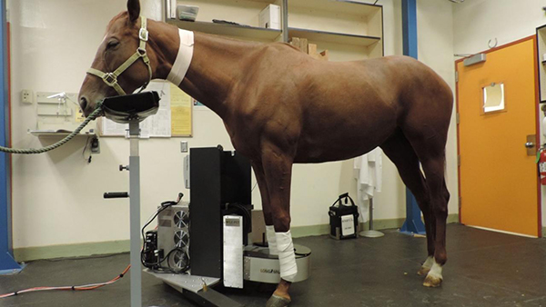
First Standing Equine PET Scanner Now Ready For Clinical Use

UC Davis
By: Rob Warren
The UC Davis School of Veterinary Medicine, in collaboration with LONGMILE Veterinary Imaging, has completed the first phase of the validation of the MILE-PET, the first positron emission tomography (PET) scanner specifically designed to image the limbs of standing horses, using light sedation, eliminating the need for anesthesia.
The first phase of this study, funded by the Grayson Jockey Club Research Foundation and conducted under the supervision of Dr. Mathieu Spriet, associate professor of veterinary radiology at UC Davis, consisted of validating the safety of the system and establishing scanning protocols using research horses from the UC Davis Center for Equine Health. Six horses were imaged twice with the standing scanner and once under general anesthesia. This allowed the researchers to confirm the repeatability of findings and to compare with results obtained with the technique previously developed on anesthetized horses.
The horses tolerated all of the procedures well. All imaging sessions were successful, and no complications were reported. The quality of images obtained on the standing horses were similar to the ones performed under general anesthesia.
“I am very excited to report that everything worked according to plan, if not better! I am very impressed with the quality of images we were able to obtain,” said Dr. Spriet.
Scan lengths ranging from 1 to 10 minutes were compared, and the team of experts concluded that a 4-minute scan is long enough to obtain images of high diagnostic quality. The rapidity of acquisition is a great advantage for clinical patients, as multiple areas can be imaged with just a short sedation time. The focus of this initial validation study has been on the fetlock joint, as this is the area most commonly injured in racehorses, but the researchers were also able to obtain high quality images of the foot and carpus (horse knee).
The MILE-PET is now ready to image racehorses in training. This clinical trial will begin at UC Davis before the scanner be moved to Santa Anita Park in mid-December. The Stronach Group, owners of Santa Anita Park, and the Southern California Equine Foundation, who operate the veterinary hospital at Santa Anita, have been key partners in the project by supporting the scanner development costs. Although this project has been in the works for more than a year, the recent highly mediatized horse fatalities in Southern California have highlighted the need for improved safety in horse racing. Availability of imaging techniques that are able to detect bone changes that might predispose to catastrophic breakdowns is one of the measures that has been proposed to reduce the track fatalities.
“PET has a very interesting role to play in racehorses, as it detects changes at the molecular level, before structural changes occur,” explained Spriet. “In other words, PET provides warning signs that injuries might happen. There is still a lot of work ahead of us, as we need to learn to distinguish the PET changes that reflect normal adaptation to speed work from changes that are indicative of high risk for major injuries.”
The plan is to image as many horses as possible at Santa Anita over the coming year. Once the researchers have established a large database representing the different patterns of PET findings in racehorses, patterns at risk for breakdown will be identified. PET will ultimately become an additional tool to help in the management of racehorses with gait abnormalities in order to prevent breakdown.


