MRI Was Key to Diagnosing Horse’s Mysterious Lameness…
Penn Vet Extra By: Louisa Shepard
The farrier did her feet that week, and Legato, known as Lexi, improved. MacGuinness chalked it up to a shoeing imbalance, but gave the big Warmblood mare the winter off, just in case.
“I checked her now and then, circling to the right, and she seemed fine, so I thought it was one and done,” MacGuinness said. “When I started riding her again in the spring, she seemed fine at first, but then all of a sudden she started to look lame to the right.“
Again, the farrier came out, and Lexi improved. But then she rapidly deteriorated, lame not just on the right turn, but also while going straight, even at a walk.
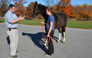
MacGuinness in April brought Lexi to New Bolton Center where she works as a patient care technician on the barn nursing staff. “I bring my horses to New Bolton because I know they will get the best of care,” she said.
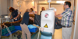
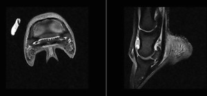
Radiographs didn’t show anything remarkable in the right front foot, said Dr. David Levine, Assistant Professor of Large Animal Surgery. He recommended a scan with New Bolton Center’s new MRI system, designed specifically for obtaining high-quality images in horses. Lexi was the first patient to use the new system, which arrived in June.
Magnetic resonance imaging (MRI) is used primarily for soft-tissue injuries associated with lameness, but it also detects bone injury. [Click here to learn more about New Bolton Center’s MRI service.]
“We diagnosed a condition using the MRI that we could not otherwise have diagnosed in that part of the foot,” Levine said.
The diagnosis: Adhesions of the deep-digital-flexor tendon to the navicular bone in the right front foot. The solution: minimally invasive surgery, a “navicular bursoscopy,” to break down the adhesions.
The navicular bone aids in the gliding of the deep-digital-flexor tendon over the coffin joint (distal-interphalangeal joint) in the foot, Levine said. “It is a very important area, and a very frequent cause of lameness that can be difficult to diagnose without MRI,” he said.
Getting a diagnosis was helpful, MacGuinness said, so she could make an informed decision on how to best treat Lexi, rather than just putting her on stall rest and hoping the lameness would resolve.
“The MRI was incredibly useful,” MacGuiness said. “Without it, we probably still wouldn’t know what was going on in there.”
Dr. Levine performed the surgery in September. “We were able to give Caitlin a diagnosis and prognosis, and an option for treatment,” he said. “I gave her a realistic outlook on what her horse’s future would be, and we performed surgery to address the problem.”
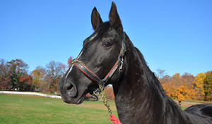
MacGuiness is an experienced dressage rider and trainer, and currently owns four horses, in partnership with her mother and aunt. Lexi was a dressage prospect, chosen for her willing nature, smooth ride, and 16.3-hand frame. The Saddlebred/Dutch Warmblood-cross has a stunning presentation – super-dark bay, almost black, with four dramatic white socks.
A pre-purchase exam was clean, and she was in full training for nearly a year when she came up lame, MacGuinness said. The cause of her injury remains a mystery.
And Lexi’s future is still uncertain. MacGuinness brought her back to New Bolton Center the first week of November for a recheck. Levine injected the distal-interphalangeal joint with an anti-inflammatory, and sent her back home for continued rehabilitation.
“The prognosis is still guarded,” MacGuiness said. “At least with the surgery she has a chance, because without it, she wouldn’t have.”


