Mare’s Sight Restored With Novel Corneal Edema Surgery
By: Sacha Adorno
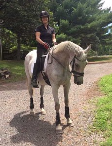
Thoroughbred mare, Set Chivalry, known to all who love her as Chevy, is a large personality. “She’s so smart and has such a spicy attitude,” said owner Susan Lax of the 21-year-old retired Dressage horse. “She’s just really the apple of my eye and so exhilarating to ride. I have had her since she was four, and she is a member of our family. I would do whatever is necessary to keep her healthy.”
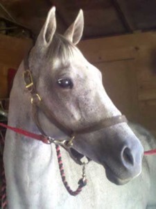
Two years ago, Chevy developed glaucoma in her left eye. Local equine ophthalmologists were unable to fix the condition. Then last year, Susan noticed Chevy developing a similar cloudiness in her right eye. She decided to make the nearly three-hour trip from her Hunterdon County, NJ, home to New Bolton Center, where Dr. Nicole Scherrer observed corneal cloudiness and diagnosed her with a painful condition called corneal edema.
In a healthy eye, the clear outer portion of the eye is the cornea and this structure stays transparent because it is dehydrated. The cornea’s endothelium, its innermost layer, plays a large role in maintaining dehydration by preventing water from filling the cornea.
The endothelium can be damaged by primary endothelial disease, trauma, uveitis, or glaucoma. When it is damaged or diseased, endothelial cells don’t work correctly, and the cornea fills with water and swells, giving the characteristic blue color and resulting in reduced vision and pain.
Saving Chevy’s Eye
“Chevy came to us with chronic eye issues,” explained Scherrer, Clinical Assistant Professor of ophthalmology at New Bolton Center. “We’d hoped to save both eyes, but her left eye was completely blind when I initially met her. All we could do was keep her comfortable. However, I felt confident that we could stop the disease progression in the right eye and save her vision.”
Scherrer admitted Chevy for monitoring and emergency surgery. In the operating room, the surgical team used an approach Scherrer developed and first tried at Penn Vet six years ago with Dr. Mary Lassaline, then on faculty at New Bolton Center and now at UC Davis School of Veterinary Medicine.
The procedure involves a superficial keratectomy, which removes a piece of the cornea. The missing piece is then replaced with a graft—or Gundersen inlay flap—of conjunctiva, pink tissue surrounding the eye.
“The conjunctiva acts as a sponge and pulls water out of the cornea,” explained Scherrer. “We cannot fix the endothelium, but we can compensate for it. Now, whenever water comes into the cornea, the graft sucks it out.”
After the procedure, the mare’s eye slowly became clear again. Scherrer said, “Eight months post-op, Chevy is doing really well at home.” She receives medication two times a day, down from six times pre-surgery, to keep swelling down.
Susan is thankful Chevy, her kindred spirit, is back to enjoying their spritely rides. “I’ve been in the horse business for more years than I’m going to admit,” remarked Susan, who owns Chivalry Hill Farm, where she trains and boards horses. “Dr. Scherrer is amazing. I text eye pictures or questions to her, and she responds quickly with advice or to allay my fears. I’ve developed an amazing relationship with her and the New Bolton Center team.”
 Novel Approach to Equine Corneal Edema
Just a few years ago, surgery wouldn’t have been an option for Chevy. Neither corneal edema nor the underlying endothelial disease respond well to medication. Historically, veterinarians have treated equine patients for pain and comfort management but not to forestall or reverse the disease.
Then in 2012, one of Scherrer’s patients inspired her to look beyond medication for an eye-saving solution.
“The owners wanted to save their horse’s eye at any cost, and medicine wasn’t helping,” said Scherrer. “We considered surgery but didn’t have a tested routine to use. Reading through old research, we came across a graft used by ophthalmologist Dr. Trygve Gundersen more than 60 years ago for corneal disease in humans. We didn’t know if it would work.”
Scherrer and Lassaline tried the superficial keratectomy and Gundersen inlay flap method, named for the graft’s developer, for the first time and saved the patient’s eye. Finding the technique successful, they began using it with other horses with early stage endothelial disease. Today, Scherrer uses it in five-10 cases per year.
In 2017, five years after the initial surgery, she was lead researcher on a paper published in Veterinary Ophthalmology about four equine corneal edema cases treated using the approach.
The report—Corneal Edema in Four Horses Treated with a Superficial Keratectomy and Gundersen Inlay Flap—found that 15 months after their procedures, the horses showed a significant decrease in corneal edema, as well as an increase in comfort levels. The treatment also eliminated corneal ulcers, a common side effect of the condition. And, even more significantly, the research reported that all four horses retained their postoperative sight without numerous medications.
Said Scherrer, “I’m hoping, with publication of this paper and by getting the word out to other veterinarians, equine ophthalmologists, and owners, that more surgeons will adopt the technique… and more horses will keep their visual eyes.”


