


Hoof Wall of the Horse


Image credit: AAEP.
By Brian S. Burks, DVM, Diplomate, ABVP, Board-Certified Equine Specialist
The hoof is a modified skin (epidermis) covering the third phalanx and all enclosed structures. It provides protection to the distal limb, formed by keratinization of the epithelial layer and modification of the underlying dermis. The thickened, cornified keratin of the epidermis is referred to as horn and makes up the outer surface of the hoof, and is resistant to mechanical and chemical damage.
Each epidermal region of the hoof is associated with a dermal region (corium). The corium are connected to the underlying structures by the subcutis. The foot can be divided into 5 segments – Wall, coronary, periople, sole, and frog – and there are 5 corresponding underlying corium.
The hoof is a form of protection to the distal phalanx and also acts as a shock absorber, reducing the concussion of impact on the structures of the distal limb. The wall of the hoof is the visible portion of the hoof, forming medial lateral, and dorsal aspects, being further divided into the toe, quarters, and heels. At the heels, the walls reflects back upon themselves at the angles to become the bars, which progress cranially along the edge of the frog. The wall is widest at the distal aspect of the hoof (toe) and decreases in width around the quarters. The wall of the hoof is 5-10mm thick and consists of three layers.
The periople extends almost 19–25 mm (¾ to 1 inch) at the top of the wall to protect the sensitive coronary band at the junction of the skin and the hoof. It is very tough and of a similar consistency to the frog. After extending nearly 20 mm (¾ inch), the periople changes into a thin layer of material that covers the rest of the hoof and gives it a shiny appearance, acting as a protective covering. It helps to control evaporation of moisture from the wall. In the early stages, this horn material is quite soft – deliberately so because it helps to prevent the coronet band becoming bruised as shock is transferred upwards through the hoof wall during the weight bearing phase of the stride. The periople covers this horn to provide protection.
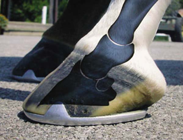
Image credit – Fox Run Equine Center.
The middle layer is the main structure of the wall, composed of amorphous horn, reinforced with tubular shaped horn rods. Its main purpose is to bear the weight of the horse. The outer hoof wall is pigmented and quite strong, protecting the inner structures of the hoof from shock and to store energy, which is released during different phases of stride to help propel the horse forward. It will be nearly impermeable, keeping water out, unless it becomes damaged via injury or nutritional imbalance.
The inner laminar layer is composed of interdigitating laminae of horn and dermal laminae which ensure that the hoof wall is well attached to the coffin bone. It is more pliable than the outer wall due to it having a higher moisture content which enables the inner wall to stretch more as the outer wall moves, ensuring the inner workings of the hoof are protected from too much shock as well as allowing the pedal bone and the outer wall to move in different ways without losing strength of attachment. This living bond between the horn (insensitive laminae) and dermis (sensitive laminae) gradually allows the hoof wall to slide distally where the distal border is worn away by ground contact.
The laminar corium consists of laminae engorged with blood vessels. The sensitive laminae mesh with the insensitive laminae of the wall on one side and are firmly attached to the pedal bone on the other side. This bond with the insensitive laminae is disrupted during laminitis. The laminar corium suspends the entire weight of the animal by the distal phalanx within the hoof capsule. Damage to the vasculature of the laminar corium can result in compromises in the integrity of the interdigitations.
The coronet band is the “crown”. Sometimes called the coronary band, with coronary pertaining to the heart. The coronary band is the primary source of growth and nutrition for most of the hoof wall. Injuries to this structure are serious and usually leave a permanent defect in the wall as it grows.
The wall of the hoof grows from the coronary band at a rate of 6-9mm (¼ to ½ inch) per month. The average toe is 76–100 mm (2½ to 4 inches) long at the toe, this means that the horse grows a new hoof in about a year. The hoof wall is made of a tough material called keratin that has a low moisture content (approximately 25% water), making it very hard and rough.
The main function of the hoof wall is to bear weight (NOT the sole). It must be trimmed at regular intervals to keep it properly shaped and level. The heel is the most elastic part of the hoof, allowing the hoof to expand during movement. Because the heel is somewhat softer than the toe, care must be taken when trimming the heel.
Content from: